Roger D Metcalf DDS, JD
Forensic Odontology
and Some Other Stuff
Odontologists--remember we need basic, foundational research in all areas of our discipline. Are our procedures scientifically valid and reliable?
Critically examine everything we've been taught. Question the scientific basis of every standard, guideline, best practice, or principle followed.
Keep in mind the quote often attributed to W. Edwards Deming: "Without data you are just another person with an opinion." I would add, if you use incorrect data, you may commit forensic malpractice, and, always remember: "First, do no harm."
We insist on evidence-based treatment in health care, why not in forensics?
Merely saying "we're following the science" without verifying that the "science" being followed is actually true is the same thing religions and cults do.
Striae of Retzius and Perikymata
Scanning Electron Microscopy
One class I took in graduate school at the University of Texas at Arlington that I really, really enjoyed very, very much was in Scanning Electron Microscopy under my major professor, Dr. Howard Arnott. We had to use a lot of photographic techniques in S.E.M. back in the day, because the images were captured on 4" x 5" film and not digitally as is done now. But I've always really enjoyed darkroom work very much, so that was ok.
When I was in this graduate program about a hundred years ago (actually, more like about 30--it was in a PREVIOUS CENTURY!), I did a little research on the anatomical structures of teeth that are called Perikymata and Striae of Retzius. Perikymata are wavy lines on the surface of teeth, and Striae of Retzius could be likened somewhat to “growth rings” inside the teeth and are seen in sagittal section. It had been speculated for some time that these two structures were actually the same anatomical feature, but just viewed from different angles—one is seen on the surface of the teeth, and one is seen when teeth are sectioned.
Being a dentist, I was very interested in things like that, and, when I took a class in graduate school on scanning electron microscopy (S.E.M.), some of the first things I looked at under the scope were teeth, naturally. After a couple of semesters I worked up a paper, and took a bunch of S.E.M. photos of some teeth and I was the first to demonstrate that Perikymata and Striae of Retzius were, in fact, continuous and were the same structure as had been speculated—the Perikymata are simply the external manifestation of the Striae.
I did this by fracturing the crowns of human teeth lengthwise and then focusing in on the sharp “corner” where external surface of the tooth and the sagittal-section of the internal part of the tooth intersected (see images at bottom of page). It was pretty cool, I thought. Now, again, this was about 30 years ago when I was a graduate student and a novice at S.E.M., not at all a professional microscopist, and working on an ancient electron microscope that was an antique even back then.
I wrote this up for Dr. Howard Arnott, my major professor in the PhD program in Quantitative Biology at the University of Texas at Arlington in 1992, and presented this at an S.E.M. Society meeting, but never submitted it for publication anywhere. I should, I guess.
Perikymata, again, are the wavy lines on the surface of teeth, and the Striae are sort of like growth rings. I wonder if they’re in individual patterns, and wonder if we can get impressions of them somehow?—maybe we could use them for identification purposes in some way.
Initially I arbitrarily selected extracted human incisors, froze them in liquid nitrogen, and carefully fractured them with a surgical mallet. Eventually I found that just smacking them at room temperature with a regular carpenter's hammer worked just as well. I would try to obtain a fracture that ran lengthwise nominally parallel to the long axis of the tooth in the sagittal plane. I then dipped the crown of the tooth in 30% HCl for about 3-5 sec and rinsed with water. The tooth was next air-dried and sputter coated with platinum/palladium in argon and the selected "corner" was examined in the S.E.M.
I also took a class in Transmission Electron Microscopy (T.E.M.) where we did electron microscopy on automobile transmissions. No, that's an old, bad joke. Now, 'scanning' electron microscopy (S.E.M.) is where one examines the external surface of some object--scanning the surface with the electron probe. Think of S.E.M. as using an ultra-powerful magnifying glass to look at the surface of the object.
'Transmission' electron microscopy, on the other hand, is where one looks through a thin section of some object, very much like when looking at slides in conventional light microscopy. Preparing the specimens for T.E.M. is exceedingly exacting, boring, difficult, tedious, and time consuming. Plus--dangerous--you have to use some pretty noxious chemicals, such as uranium and osmium compounds, to stain the sections of biological specimens. U.T. Arlington has a very active materials science department, and they used the T.E.M.s most of the time, and the biology department didn't really do all that much T.E.M. at U.T.A.--at least, not when I was there. Conversely, materials science at U.T.A. didn't do so much S.E.M., but biology used the S.E.M.'s a lot. We have two brand-new S.E.M.'s at the office and the trace evidence folks use them a LOT. I hope to convince 'em to let me get some time on the new S.E.M.'s.
The images below are images of the fracture line where the external surface of a human tooth meets the inner enamel sagittal-section. These probably don't mean too much unless you are really interested in teeth!
The images below are the first known S.E.M. images to show that Perikymata are the external manifestation of the Striae of Retzius.
Images (C) 1992, 1993, 2003, and 2013 Roger D Metcalf DDS. All rights reserved, worldwide. No reproduction without permission. All images are digitally registered and watermarked.
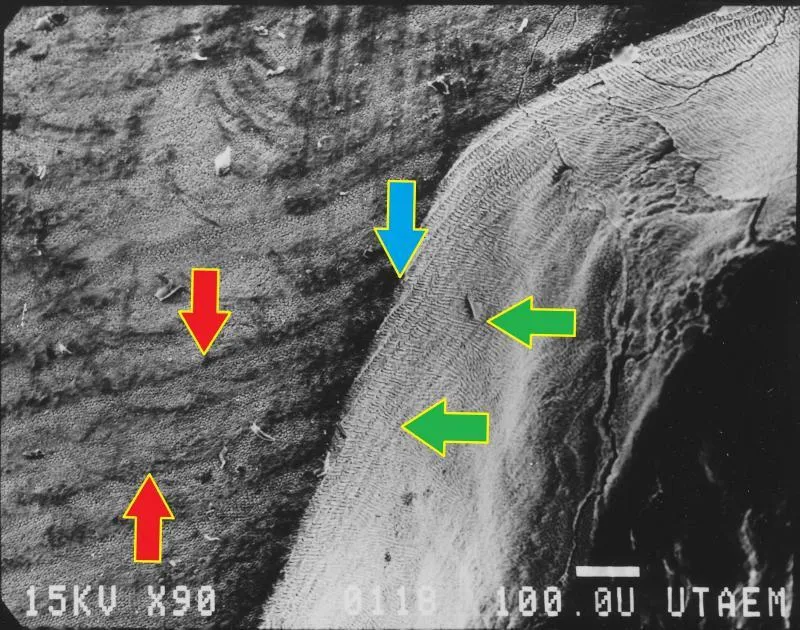
Above: a 90x view of a tooth. The red arrows point to the wavy lines on the external enamel surface which are the Perikymata.
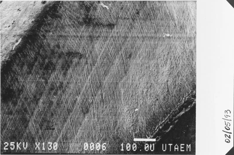
Above: 130x oblique view of the interior of the tooth in sagittal plane.
Above: 220x view of the fracture line. On the L are the Perikymata and on the R are the Striae of Retzius.
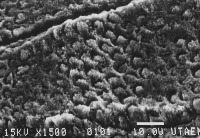
Below: etched human dentin, 1500x
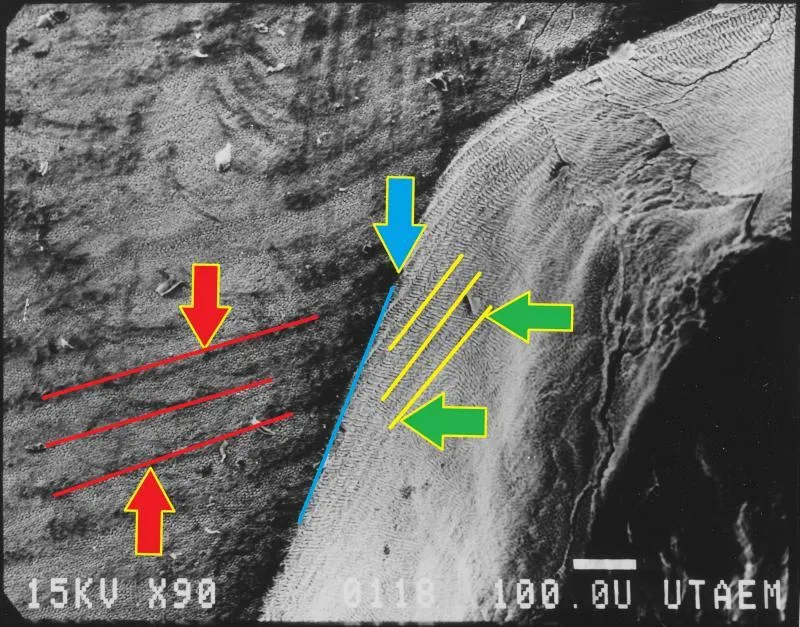
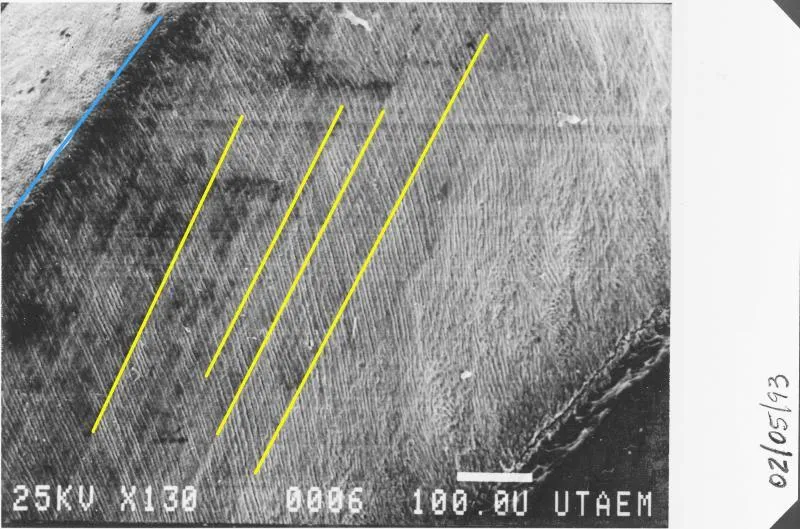
Above: 90x view. The blue arrow points to the fracture line where the external surface of the enamel (L) intersects with the interior of the tooth (R) in sagittal section. The yellow lines indicate the Striae of Retzius.
Above: 220x view of the fracture line. On the L are the Perikymata and on the R are the Striae of Retzius.

Above: and again, the red lines indicate the Perikymata on the surface of the enamel (L), and the yellow lines indicate the Striae of Retzius in the sagittal plane (R); the blue line is the "corner" of the intersection.
Below: etched enamel surface, if you ever wanted to see human enamel prisms, 1500x.
Roger D Metcalf DDS, JD
PO Box 137442
Fort Worth, TX 76136-1442
ph: +1-817-371-3312
fax:+1-817-378-4882
© 2013 - 2024. Roger D Metcalf. All worldwide rights reserved.
No reproduction without permission.
Neither the Tarrant County Medical Examiner's District, Tarrant County, the American Board of Forensic Odontolgy, the American Society of Forensic Odontology, the Royal College of Physicians, Oklahoma State University, nor any other organizaion mentioned here necessarily supports or endorses any information on this website. Any opinions, errors, or omissions are my responsibility, and mine alone. This site DOES NOT REPRESENT the official views of any of these--or any other-- organizations. Similarly, those other organizations may not fully represent my views, either.